561.391.1728


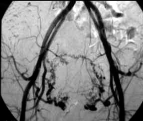
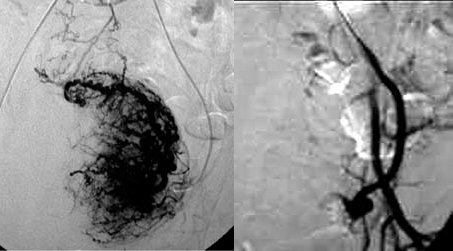
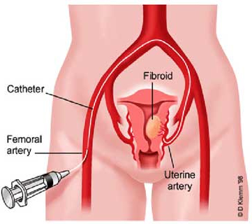
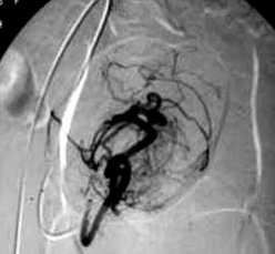
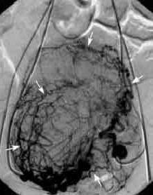
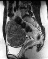
Injection of the left uterine artery shows enlarged, tortuous arteries supplying the fibroids.
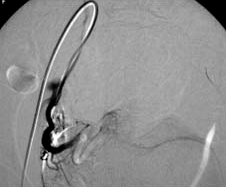
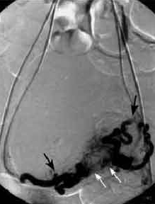
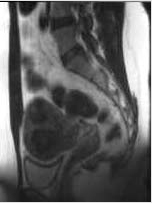
Cessation of blood flow forward to the fibroids, with reflux of contrast to arteries in posterior division of the left internal iliac artery following embolization.
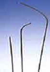
 Angiographic
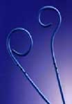
 Catheters
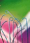
 Guidewires
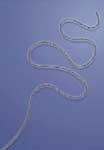 Microwires
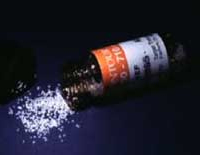
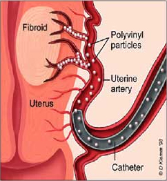
Polyvinyl particles
Polyvinyl particles (PVA), used to embollize the arteries that supply the fibroids are tiny synthetic "plastic" particles that are the size of fine grains of sand ranging from 0.3 mm to 0.7 mm in diameter. PVA has been used for more than 20 years in embolization of other parts of the body with no known reports of any adverse side effects due to this embolization material.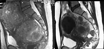
BOCA RATON REGIONAL HOSPITAL
DEPARTMENT OF VASCULAR
& INTERVENTIONAL RADIOLOGY
800 Meadows Road
Boca Raton, Florida USA 33486
 Free counters provided by

ADA Compliance Policy: In accordance with the American with Disabilities Act, Boca Radiology Group strives to ensure that our web site is accessible to individuals with disabilities. If you have any questions or suggestions regarding the accessibility of this site, please contact us via email or call us at 561.391.1728, so we can ensure you receive the information you are seeking.
© 2023 All Rights Reserved